Upper Leg Tendon Anatomy - Upper Legs And Psoas Muscles Anatomy Stock Photo Picture And Royalty Free Image Image 89697634. This is why you have to indicate which biceps you are taking about when discussing one or other of these muscles. Upper leg anatomy and function the upper leg is often called the thigh. On the medial edge of the posterior thigh is the gracilis muscle. It is the junction of the thigh and the leg and is a hinge joint. The quadriceps tendon attaches the quadriceps muscles to the patella.
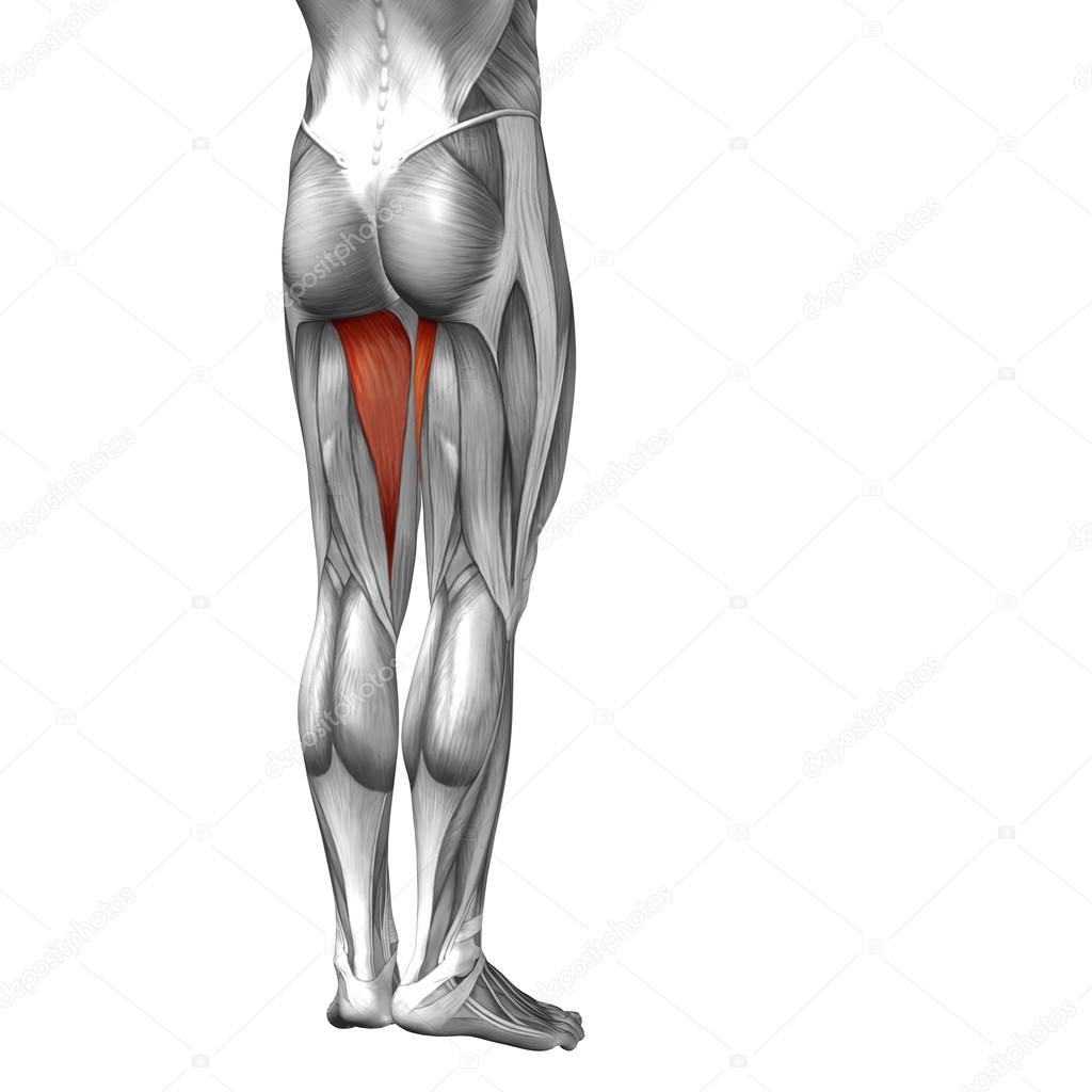
The patella is attached to the shinbone (tibia) by the patellar tendon. Possibly the most important tendon in terms of mobility is the achilles tendon. This tendon can get irritated from overuse, muscle weakness and muscle tightness. The human leg, in the general word sense, is the entire lower limb of the human body, including the foot, thigh and even the hip or gluteal region. 430) is the most superficial muscle on the medial side of the thigh.
Conceptual 3d Human Upper Leg Anatomy Or Anatomical And Muscle Isolated On White Stock Photo Image By C Design36 94597430 from st2.depositphotos.com 430) is the most superficial muscle on the medial side of the thigh. It is the junction of the thigh and the leg and is a hinge joint. Upper leg tendon anatomy this mri wrist coronal cross sectional anatomy tool is absolutely free to use. Muscle, organ and skeletal anatomy). The vastus medialis (vastus medialis oblique, or vmo) is one of the four quadriceps muscles in the front of your upper thigh. Related online courses on physioplus. Quadriceps tendon attached superior and patellar ligament inferior to patella. Tenderness to the touch at the front of your hip;
It is the junction of the thigh and the leg and is a hinge joint.
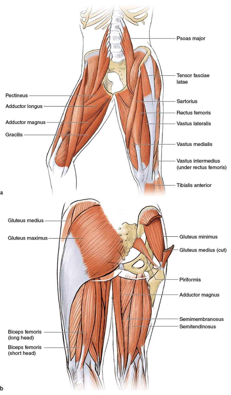
Upper leg anatomy and function the upper leg is often called the thigh. The detailing of these structures changes based on dog breed due to the huge variation of size in dog breeds. The knee joint is commonly injured, so understanding its anatomy can help you understand the conditions that cause problems, so you stay safe and prepared. They consist of the rectus femoris, vastus intermedius, vastus lateralis and the vastus medialis. Tendons are thick bands of tissue that connect muscles to bone. Other muscles of the anterior (front) thigh include the pectineus, sartorius,. 430) is the most superficial muscle on the medial side of the thigh. This is why you have to indicate which biceps you are taking about when discussing one or other of these muscles. We speak of the upper extremities (arms) and the lower extremities (legs). Squeeze your knees together and boom, you're contracting the adductors. It's the area that runs from the hip to the knee in each leg. Its muscle belly is on the back aspect of the upper arm. Related posts of muscle anatomy upper leg.
It's the area that runs from the hip to the knee in each leg. Lateral (fibular) collateral ligament (fcl) upper part middle part lower part popliteus tendon (pt) upper part i. It is thin and flattened, broad above, narrow and tapering below. Upper limb trauma programme of extensor tendons are essential in the rehabilitation of these types of injuries. It serves to attach the plantaris, gastrocnemius (calf) and soleus muscles to the calcaneus (heel) bone.
Upper Legs Running Anatomy Sports Anatomy from doctorlib.info Possibly the most important tendon in terms of mobility is the achilles tendon. Muscle anatomy of upper thigh. These muscles run from the lower spine and pelvis, join together, then attach by a tendon to the upper thigh. The human leg, in the general word sense, is the entire lower limb of the human body, including the foot, thigh and even the hip or gluteal region. Medial muscles adduct and rotate your thigh, and posterior flex your leg and extend your thigh. Anatomically speaking, your thigh is the area of your upper leg between your hip joint and your knee. Its muscle belly is on the back aspect of the upper arm. In clinical anatomy the thigh muscles are divided into three groups:
Muscle anatomy of upper thigh.
They consist of the rectus femoris, vastus intermedius, vastus lateralis and the vastus medialis. Dog anatomy details the various structures of canines (e.g. Upper leg tendon anatomy : Upper leg tendon anatomy from i0.wp.com the achilles tendon or heel cord, also known as the calcaneal tendon, is a tendon at the back of the lower leg, and is the. The muscles of the leg anatomy chart shows in every possible view the way that the muscles and other pieces of the leg work together in motion and flexibility. On the medial edge of the posterior thigh is the gracilis muscle. Upper leg tendon anatomy : Medial muscles adduct and rotate your thigh, and posterior flex your leg and extend your thigh. These muscles run from the lower spine and pelvis, join together, then attach by a tendon to the upper thigh. The sartorius is known for two types of pain, burning stinging sensations and sharp stabbing pain. Lateral (fibular) collateral ligament (fcl) upper part middle part lower part popliteus tendon (pt) upper part i. The vastus medialis (vastus medialis oblique, or vmo) is one of the four quadriceps muscles in the front of your upper thigh. They bear the weight of the upper body.
We speak of the upper extremities (arms) and the lower extremities (legs). Muscle, organ and skeletal anatomy). Upper leg tendon anatomy : Muscle anatomy coloring book 12 photos of the muscle anatomy coloring book anatomy coloring book muscles free, muscle anatomy coloring book, muscle anatomy coloring book pdf, muscle anatomy coloring pages free, muscular anatomy coloring book, human muscles, anatomy coloring book muscles free, muscle anatomy. Upper leg tendon anatomy from i0.wp.com the achilles tendon or heel cord, also known as the calcaneal tendon, is a tendon at the back of the lower leg, and is the.
0514 Upper Legs Anterior View Medical Images For Powerpoint Powerpoint Templates Download Ppt Background Template Graphics Presentation from www.slideteam.net Squeeze your knees together and boom, you're contracting the adductors. We speak of the upper extremities (arms) and the lower extremities (legs). Human muscles · july 20, 2016. It is also visible on the medial edge of the thigh from the anterior. In clinical anatomy the thigh muscles are divided into three groups: It's the area that runs from the hip to the knee in each leg. The burning and stinging sensations are associated with trigger points in the muscle. The fibers run vertically downward, and end in a rounded tendon, which passes behind the medial condyle.
Upper leg tendon anatomy from i0.wp.com the achilles tendon or heel cord, also known as the calcaneal tendon, is a tendon at the back of the lower leg, and is the.
One more example is the large muscle group of the quadriceps, located on the front of the upper leg. This tendon can get irritated from overuse, muscle weakness and muscle tightness. Anatomically speaking, your thigh is the area of your upper leg between your hip joint and your knee. Sometimes thigh pain can occur after trauma or an injury, and other times it may come on for no apparent reason. These muscles run from the lower spine and pelvis, join together, then attach by a tendon to the upper thigh. 430) is the most superficial muscle on the medial side of the thigh. The fibers run vertically downward, and end in a rounded tendon, which passes behind the medial condyle. Anterior muscles extend your legs and flex your thighs. Your upper leg includes seven major muscles. Upper leg anatomy and function the upper leg is often called the thigh. Other muscles of the anterior (front) thigh include the pectineus, sartorius,. Tendons are cords made of tough tissue, and they work as special connector pieces between bone and muscle. This is why you have to indicate which biceps you are taking about when discussing one or other of these muscles.